To
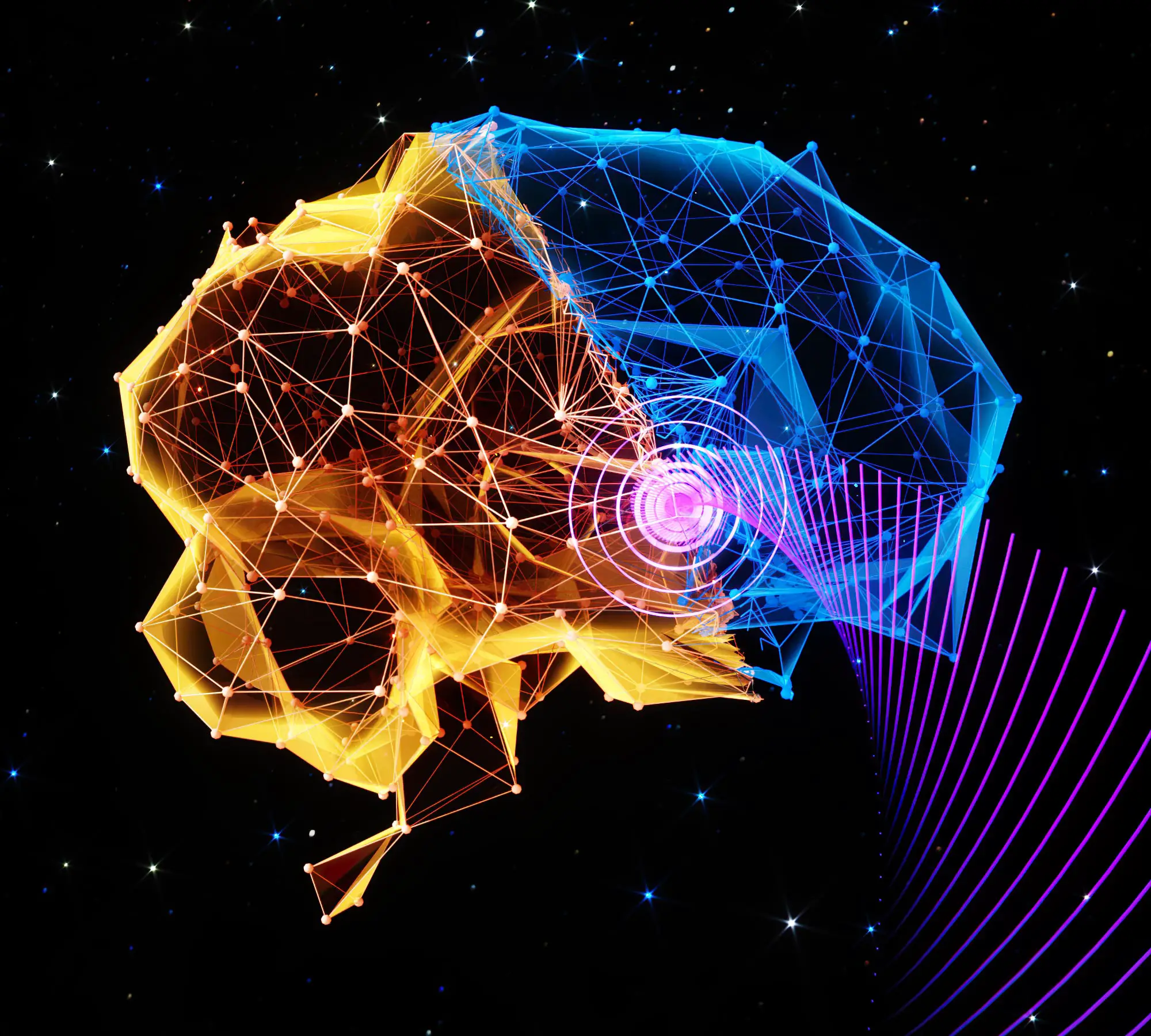
A multidisciplinary team led by Associate Professor Hong Cheng of Washington University in St. Louis has developed a new non-invasive method of inducing anesthesia in mammals by targeting the central nervous system with ultrasound. This technique of stimulating the preoptic area of the brain’s hypothalamus was shown to effectively lower body temperature and metabolic rate in mice, leading to numbness. This is a natural mechanism some animals use to survive extreme conditions.Credits: Image courtesy Chen Lab, University of Washington, St. Louis
Scientists at the University of Washington in St. Louis have developed a method to induce lethargy in mammals using ultrasound stimulation of the brain, according to a study. natural metabolism. This non-invasive technique could potentially be used in scenarios such as spaceflight and to conserve energy and heat in patients with severe health conditions.
Some mammals and birds have clever ways of conserving energy and heat by going into a coma. In this state, their body temperature and metabolic rate are lowered, allowing them to survive in potentially lethal environmental conditions such as extreme cold and lack of food. Similar conditions were proposed for spaceflight scientists in the 1960s and for patients with life-threatening health conditions, but such conditions remain difficult to induce safely.
Hong Cheng, an associate professor at Washington University in St. Louis, and his multidisciplinary team induced lethargy in mice by ultrasonically stimulating the hypothalamic preoptic area of the brain, which helps regulate body temperature and metabolism. In addition to mice that spontaneously fall into a coma, Cheng and her team also induced coma in rats, which did not go into a coma.Their findings were published in the journal May 25. natural metabolismdemonstrate the first non-invasive and safe method to induce a numb-like state by targeting the central nervous system.
Cheng’s team used ultrasound to safely and noninvasively induce lethargy-like states in mice and rats.Credits: Video Courtesy: Chen Lab, University of Washington, St. Louis
Chen, associate professor of biomedical engineering at the McElvey School of Engineering and radiation oncology at the School of Medicine, and her team, which includes postdoctoral fellow Yaoheng (Mak) Yang, created a wearable ultrasound transducer that stimulates neurons in the body. hypothalamic preoptic area.When stimulated, the body temperature of the mice dropped by about 3 degrees[{” attribute=””>Celsius for about one hour. In addition, the mice’s metabolism showed a change from using both carbohydrates and fat for energy to only fat, a key feature of torpor, and their heart rates fell by about 47%, all while at room temperature.
The team also found that as the acoustic pressure and duration of the ultrasound increased, so did the depth of the lower body temperature and slower metabolism, known as ultrasound-induced hypothermia and hypometabolism (UIH).
“We developed an automatic closed-loop feedback controller to achieve long-duration and stable ultrasound-induced hypothermia and hypometabolism by controlling of the ultrasound output,” Chen said. “The closed-loop feedback controller set the desired body temperature to be lower than 34 C, which was previously reported as critical for natural torpor in mice. This feedback-controlled UIH kept the mouse body temperature at 32.95 C for about 24 hours and recovered to normal temperature after ultrasound was off.”
To learn how ultrasound-induced hypothermia and hypometabolism is activated, the team studied the dynamics of the activity of neurons in the hypothalamus preoptic area in response to ultrasound. They observed a consistent increase in neuronal activity in response to each ultrasound pulse, which aligned with the changes in body temperature in the mice.
“These findings revealed that UIH was evoked by ultrasound activation of hypothalamus preoptic area neurons,” Yang said. “Our finding that transcranial stimulation of the hypothalamus preoptic area was sufficient to induce UIH revealed the critical role of this area in orchestrating a torpor-like state in mice.”
Chen and her team also wanted to find the molecule that allowed these neurons to activate with ultrasound. Through genetic sequencing, they found that ultrasound activated the TRPM2 ion channel in the hypothalamus preoptic area neurons. In a variety of experiments, they showed that TRPM2 is an ultrasound-sensitive ion channel and contributed to the induction of UIH.
In the rat, which does not naturally go into torpor or hibernation, the team delivered ultrasound to the hypothalamus preoptic area and found a decrease in skin temperature, particularly in the brown adipose tissue region, as well as about a 1 degree C drop in core body temperature, resembling natural torpor.
This multidisciplinary team consists of Jonathan R. Brestoff, MD, PhD, an assistant professor of pathology and immunology at the School of Medicine; Alexxai V. Kravitz, an associate professor of psychiatry, of anesthesiology and of neuroscience at the School of Medicine, and Jianmin Cui, a professor of biomedical engineering at the McKelvey School of Engineering, all at Washington University in St. Louis. The team also includes Michael R. Bruchas, a professor of anesthesiology and of pharmacology at the University of Washington.
“UIH has the potential to address the long sought-after goal of achieving noninvasive and safe induction of the torpor-like state, which has been pursued by the scientific community at least since the 1960s,” Chen said. “Ultrasound stimulation possesses a unique capability to noninvasively reach deep brain regions with high spatial and temporal precision in animal and human brains.”
Reference: “Induction of a torpor-like hypothermic and hypometabolic state in rodents by ultrasound” by Yaoheng Yang, Jinyun Yuan, Rachael L. Field, Dezhuang Ye, Zhongtao Hu, Kevin Xu, Lu Xu, Yan Gong, Yimei Yue, Alexxai V. Kravitz, Michael R. Bruchas, Jianmin Cui, Jonathan R. Brestoff and Hong Chen, 25 May 2023, Nature Metabolism.
DOI: 10.1038/s42255-023-00804-z
This work was supported by the National Institutes of Health (R01MH116981, UG3MH126861, R01EB027223, and R01EB030102). JRB is supported by NIH (DP5 OD028125) and Burroughs Wellcome Fund (CAMS #1019648).
