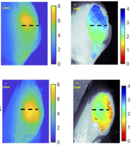
Image: Researchers have developed a system that uses dual wavelength light to provide information about the depth of tumors in healthy tissue.
look more
Credit: Christine M. O’Brien, Washington University in St. Louis
WASHINGTON — Researchers have developed a low-cost, simple imaging system that uses tumor-targeting fluorescent molecules to determine the depth of tumor cells in the body. This portable system could ultimately help surgeons distinguish between healthy and cancerous tissue with greater accuracy when resecting tumors.
Doctors use fluorescent molecules to make cancer cells glow during tumor removal, allowing the surgeon to see if any cancer tissue remains. However, the equipment required for this technique is not widely available and typically does not provide quantitative information about how deep cancer cells reside within the tissue. Having access to depth information has been shown to allow surgeons to remove a complete, healthy layer of tissue around the tumor, yielding the best possible outcome for the patient.
“The few commercial systems that provide quantitative depth information are large and expensive, limiting their use outside of large medical centers.” Washington University School of Medicine in St. Louis“Our group builds on previous work in this field to develop a low-cost, simple system that can rapidly determine tumor cell depth using near-infrared (NIR) fluorescent probes. built upon.”
Researchers describe new system in Optica Publishing Group journal Biomedical Optics ExpressThe portable, easy-to-use system can be used in resource-poor clinical centers and helps minimize health disparities.
“Such systems could be used in the future to improve surgical outcomes for patients undergoing tumor resection,” said O’Brien. “It also eliminates the need to wait for pathology results after tumor removal before seeing if cancer cells are still present.”
shedding light on cancer
Studies have shown that surgical treatment of cancer tends to be most successful when the surgeon removes not only the tumor, but also the healthy layer of tissue that completely surrounds it. However, this can be challenging as it is difficult to identify the margin between the end of the tumor and the beginning of healthy tissue. Furthermore, the optimal thickness of the normal layer depends on the tumor type and location.
To aid in this work, the Achilefu Lab research team, led by O’Brien, developed a new instrument based on the application of a single fluorescent dye during tumor resection. It is excited by two different NIR wavelengths that penetrate to different depths of tissue. His emitted NIR fluorescence can be imaged through tissue and can detect cancer cells one to two centimeters below the surface.
Dual-wavelength excitation fluorescence takes advantage of the fact that different colors or wavelengths of light travel different distances in tissue. By illuminating fluorescent tumor-targeting molecules with different wavelengths of light and comparing their responses, it is possible to predict how deep a tumor-targeting agent is located in tissue.
“Several research groups have contributed to the development of a mathematical relationship linking fluorophore depth to ratiometric fluorescence measurements,” said O’Brien. “The proliferation of near-infrared contrast agents being developed for use in medicine has allowed us to build on previous research and create a system that works in the near-infrared, is low cost, and is easy to use.”
Construction of dual wavelength system
The new fluorescence imaging system uses 730 nm and 780 nm LEDs to provide two wavelengths of excitation light and a monochrome CMOS camera to detect the resulting fluorescence. An 850 nm LED was also incorporated to create brightfield images that allow fluorescence images to be correlated with real-world views of tissue. The researchers decided to use an experimental drug called LS301. LS301 can be administered during tumor resection developed in the Atilef lab. As an infrared probe to target cancer, its broad excitation spectrum eliminates the need to use multiple fluorophores. made clinical application more complex. LS301 is currently in clinical trials in breast cancer patients.
After testing the system on layered synthetic material and chicken slices, the researchers evaluated its ability to predict tumor depth from breast tumors grown in mice. This was done by injecting mice with her LS301 and imaging them using the system. It took him five minutes to capture the required image. These image-based calculations correlated well with the true depth of the tumor, with an average error of only 0.34 mm, and may be suitable for clinical use.
Researchers are now working to make the system even more useful for surgical guidance by speeding up data processing and adding automation to the system so that it can scan entire tissue surfaces.
paper: CM O’Brien, K. Bishop, H. Zhang, X. Xu, L. Shmuylovich, E. Conley, K. Nwosu, K. Duncan, SB Mondal, G. Sudlow, S. Achilefu, “Quantitative tumor depth Using dual-wavelength excitation fluorescence” Biomed. option. express, Volume 13, Issue 1qq, pp. 5628-5642 (2022).
Doi: https://doi.org/10.1364/BOE.468059
About Biomedical Optics Express
Biomedical Optics Express provides the biomedical optics community with rapid, open-access, peer-reviewed articles related to optics, photonics, and imaging in biomedicine. The journal’s scope includes basic research, technology development, biomedical research, and clinical applications. It is published monthly by Optica Publishing Group and edited by his Ruikang (Ricky) Wang at the University of Washington, USA. For more information, see: Biomedical Optics Express.
About Optica Publishing Group (formerly OSA)
Optica Publishing Group A division of Optica (formerly OSA), Advancing Optics and Photonics Worldwide. We publish the largest collection of peer-reviewed content in optics and photonics, including 18 prestigious journals, leading member journals, and over 835 conference papers (including over 6,500 related videos). Optica Publishing Group searches, discovers and accesses over 400,000 journal articles, conference papers and videos representing all research in this field from around the world.
journal
Biomedical Optics Express
article title
Quantitative tumor depth measurement using dual-wavelength excitation fluorescence
Article publication date
October 6, 2022
Disclaimer: AAAS and EurekAlert! EurekAlert! is not responsible for the accuracy of news releases posted. Use of information by contributors or via the EurekAlert system.
